
Here are all 93 Physiology (with Special Reference to Mammals) questions asked in UPSC Zoology Optional Paper II from 2004 to 2026, arranged under the 8 topics of the official syllabus. Within each topic the latest questions come first.
Each question ends with a code such as [2024/15m/3b] – the year, the marks, the word limit (where UPSC set one) and the question number in the paper.
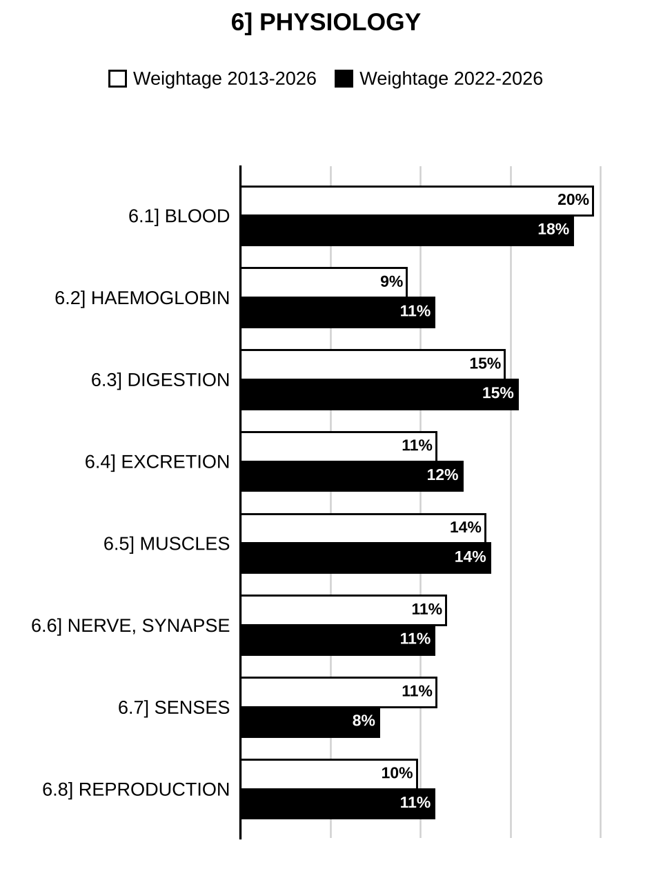
6.1] Blood
17 questions
Describe characteristic features and specific functions of lymphocytes, monocytes and neutrophils. [2025/15m/6b]
ABO system of blood groups and its basis, and erythroblastosis foetalis. [2024/10m/150w/5b]
List various coagulation factors and describe their role in blood clotting. [2023/15m/8b]
Explain the functions of neutrophils, basophils and lymphocytes. [2022/10m/150w/5c]
Define thermoregulation. How do homeotherms regulate body temperature in hot and cold climates? [2022/15m/7b]
Explain the respiratory regulation of acid-base balance. [2021/10m/150w/5c]
What is the general mechanism of blood coagulation? Describe the intrinsic pathway for the initiation of blood clotting. [2020/15m/6b]
What is meant by Rh factor? Explain Rh incompatibility in human beings. [2017/10m/5d]
Explain ABO blood groups with its genetic basis and significance in blood transfusion. [2016/10m/1b]
Elucidate the importance of thermoregulation with reference to its relationship with homeostasis in endothermic animals. [2016/20m/8a]
What is haematopoiesis? Indicate the site and the stages by which erythrocytes, various types of leycocytes and platelets are formed. Which chemical factor(s) coordinate and control these events? [2014/20m/6c]
What do you mean by anticoagulant? Describe the structure and functions of natural and synthetic anticoagulants. [2014/15m/7c]
Mention the different steps in the metabolism of iron. What are the functions that require iron? Add a note on the regulatory mechanisms that ensure stable titers of iron in the body. [2013/10m/5a]
What are erythroid cells? Explain the RBC lineage in balancing haemoglobin manufacture. [2013/15m/8a]
What controls heartbeat, and the rate at which blood flows through the circulatory system? [2012/20m/3c]
What is Rh factor and how is it determined? What is the cause and what is/are the symptom(s) of erythroblastosis fetalis, and how can it be corrected? [2012/20m/6b]
Give an account of factors and physiology of blood coagulation. [2004/60m/7]
6.2] Haemoglobin and respiratory gases
8 questions
Explain sigmoidal nature of oxygen dissociation curve for hemoglobin. [2025/10m/7a(i)]
Describe differences between adult and fetal hemoglobin and comment on their physiological significance. [2025/10m/7a(ii)]
Explain the structure of haemoglobin and its role in carbon dioxide transport. [2023/15m/6b]
Explain the transport of oxygen in blood. Discuss the factors that shift oxygen-hemoglobin dissociation curve. [2021/15m/7b]
Explain the structure and functions of haemoglobin. [2019/15m/8b]
Give a comprehensive account of gaseous transport in blood. [2017/20m/6a]
Give the chemical composition of human haemoglobin and discuss its role in transport during various physiological states. [2011/20m/2c]
Give short descriptions of any three of the following: Chloride shift. [2004/20m/5b]
6.3] Digestion and absorption
14 questions
Write about the role of liver and pancreas in digestion. Add a note on neural and hormonal regulation of their secretions. [2024/20m/8a]
Highlight role of various hormones in the regulation of digestive secretions. [2023/10m/150w/5a]
What are intestinal glands? Describe the roles of intestinal juices in digestion and absorption of food. [2022/20m/7a]
Explain the basic stimuli causing pancreatic secretion. Discuss the role of pancreas in major food digestion. [2021/20m/7a]
Enlist the composition of saliva. Describe the mechanism of secretion of saliva. [2020/10m/5c]
Tabulate and describe the digestive enzymes, their occurrence, substrates and end products. [2019/15m/7b]
Explain the role of pancreatic enzymes in digestion. [2018/10m/5d]
Describe the process of digestion and absorption in small intestine. [2017/10m/5c]
Explain the role of salivary glands, liver, pancreas and intestinal glands in fat and carbohydrate digestion. [2015/15m/8b]
Name the enzymes and hormones released by liver and pancreas. Discuss their functions in digestion of food and maintenance of blood glucose balance. [2014/10m/5b]
Compare the role of pancreatic and intestinal glandular secretions in the digestion of food. [2012/15m/1c]
Types and functions of mammalian salivary glands. [2010/15m/150w/5b]
Discuss the role of liver and pancreas in digestion? [2007/60m/8]
Explain how hormones and enzymes control the digestion of proteins, carbohydrates and fats among mammals. [2005/60m/6]
6.4] Excretion and osmoregulation
11 questions
How is excretion different from osmoregulation in mammals? [2026/10m/150w/5b]
Explain the structure of nephron and its role in urine formation. Add a note on hormonal regulation of urine formation. [2024/20m/6a]
Illustrate the counter-current mechanism by vasa recta and mention its physiological importance. [2022/10m/150w/5d]
Describe the process of glomerular filtration. Discuss the determinants of glomerular filtration rate in human. [2020/15m/8c]
Describe in detail about the structure and function of Nephron and explain fluid balance regulation through Renin-Angiotensin-Aldosterone systems. [2018/15m/8b]
Discuss the mechanism involved in the survival of estuarine animals in varying salinities. [2016/10m/5c]
Label the parts of nephron and answer the following questions: (i) Which region of the kidney contains malpighian body, loop of Henle and collecting ducts? (ii) What is Glomerular filtration rate? (iii) Write the steps of urine formation. (iv) What is the effect of vasopressin, oxytocin and aldosterone in renal function? (v) What is the holding capacity of urinary bladder in man and the term used for the discharge of urine?
[Figure: Unlabelled diagram of nephrons with their blood supply, numbered pointers 1–15 (to be named). At the left two parallel vertical blood vessels with downward arrow 14 (left vessel, flow down) and upward arrow 15 (right vessel, flow up). A horizontal dashed line across the figure marks the boundary between the outer (upper) and inner (lower) regions of the kidney. Upper (cortical) nephron: pointer 1 – the vessel branching from the left vessels towards the capillary tuft, with an arrow showing flow towards it; pointer 2 – the capillary tuft (glomerulus); pointer 3 – the cup-shaped capsule around it; pointer 4 – the vessel leaving the tuft; pointer 5 – the coiled tubule next to the capsule, covered by a network of capillaries; pointer 6 – a second coiled tubule (upper right) joining a vertical duct; pointer 7 – the point near the bottom of the coiled tubules where the tubule and a vessel meet (below the coils). A long vessel with an arrow returns to the left vessels. Pointer 8 – the vertical branched duct at the right receiving tubules. A lower nephron (near the dashed line) has its own capillary tuft and capsule, a vessel with arrows, and long hairpin loops descending below the dashed line. Pointer 9 – two leaders to the outer vessel loop surrounding the long hairpin loop; pointers 10, 11, 12 and 13 – the limbs of the long hairpin loops in the lower region (13 and 12 on the left/descending side, 11 and 10 on the right/ascending side, inner and outer).] [2015/20m/8a]
Draw a labelled diagram of a metanephric neuron to explain (i) ultrafiltration, (ii) absorption and (iii) secretion. Define the role of hormones in these processes. [2011/15m/5d]
Write briefly on the control and regulation of renal excretion with reference to mammals. [2010/20m/7b]
Describe the structure of a mammalian nephron and explain how urine is formed. [2009/30m/7b]
What do you understand by osmoregulation? Describe the phenomenon and physiology of urine formation. [2006/60m/7]

Preparing for Prelims too?
Our UPSC Prelims Topic-wise PYQ eBook covers every Prelims question with detailed explanations, so you can see exactly where UPSC asks from.
View the BookDownload Free Sample6.5] Muscles
10 questions
Role of calcium in muscle contraction. [2026/10m/150w/5a]
Name the proteins and their functions involved in skeletal muscle contraction. Unlike skeletal muscles, why do cardiac muscles not fatigue? [2026/20m/7a]
Explain the mechanism of contraction in skeletal muscle. What do you mean by Rheobase and Chronaxie? [2024/15m/7c]
Enlist the sources of energy for muscle contraction. Describe the mechanism of contraction of skeletal muscle. [2020/20m/7a]
Enlist and describe the types and mechanical properties of muscle fibres. [2019/15m/6c]
Define Muscle twitch and explain the features of Summation and Tetanus. [2018/10m/5c]
Describe the ultrastructure of ‘Sarcomere’ and explain the process of muscle contraction. [2017/15m/6b]
Explain with diagrams the molecular mechanism of contraction of skeletal muscles. Discuss the role of ions and bioenergetics in muscle contraction. Attribute the cause(s) for muscle cramps. [2015/20m/6a]
Draw a diagram to show the arrangements of various types of contractile proteins in skeletal muscles, and explain the bioenergetics of contraction. [2012/20m/4c]
Molecular mechanism of skeletal muscle contraction. [2009/20m/5c]
6.6] Neuron and synapse
14 questions
What is nerve impulse? How is the action potential generated and propagated across the nerve membrane? [2026/15m/6b]
Transmission of nerve impulse through synapse. [2025/10m/150w/5e]
List different neurotransmitters. Explain the role of specific neurotransmitter across neuromuscular junction. [2023/10m/150w/5d]
Describe the rapidly acting synaptic transmitters with suitable examples. [2021/10m/150w/5d]
Draw a typical neuron action potential. Discuss the ionic basis for the propagation of nerve impulse. [2020/10m/5b]
Physiology of action potential. [2019/10m/5d]
Describe the role of Acetylcholine in neuromuscular transmission. [2018/10m/5a]
Describe the sequence of stages during synaptic transmission with acetylcholine as the neurotransmitter. [2016/15m/7c]
What do you mean by neurotransmitters? Describe adrenergic, cholinergic and peptidergic neurotransmitters and write an illustrated account of their functions in synaptic transmission. [2014/15m/6a]
On your answer-book, write down the name of each of the ganglia marked (i) to (vii) in the diagram below on the parasympathetic and sympathetic divisions of autonomic nervous system:
[Figure: Diagram of the autonomic nervous system drawn on either side of the brain stem and spinal cord; the organs are drawn as small unlabelled sketches. Left half headed “Parasympathetic Nervous System”, right half headed “Sympathetic Nervous System”. The brain stem shows the origins of cranial nerves labelled III, VII, IX and X; the spinal cord below is divided into segments, with T1 (first thoracic), L1, L5 and S1, S2, S3, S4, S5 labelled. Left (parasympathetic) side: four ganglia drawn as circles with dashed pointers – (iii) the ganglion on nerve III supplying the eye; (ii) a ganglion on the nerve VII pathway supplying a small gland just below the eye; (i) a second ganglion on the nerve VII pathway, just below (ii), supplying a gland drawn as a solid black body; (iv) a ganglion on the nerve IX pathway supplying a gland below it. Nerve X runs down to the heart, lungs, stomach, upper gut and intestine, and fibres from sacral segments S2–S4 run to the lower gut, urinary bladder and reproductive organs, with small terminal ganglia on or near the organs (unmarked). Right (sympathetic) side: a chain of paravertebral ganglia beside the spinal cord from the neck to the sacral region, with fibres to the head, heart and lungs; three larger prevertebral ganglia receive fibres from the thoracic and lumbar chain: (v) the uppermost, supplying stomach, liver, pancreas and nearby abdominal organs; (vi) below it, supplying the intestine; (vii) the lowest, at lower lumbar level, supplying the urinary bladder and lower pelvic organs.]
How do they regulate the visceral and reflex functions of the body? [2012/15m/1b]
With suitable examples, compare the chemical structure and functions of acetylcholine (in cholinergic neuron), adrenaline (in adrenergic neuron) and serotonin/5-hydroxytryptamine/-aminobutyric acid (in peptidergic neurons). [2012/20m/6c]
Differentiate between electrical and chemical synapse. How does summation occur at the axon hillock? [2011/15m/1d]
Excitatory and inhibitory neurotransmitters. [2010/15m/150w/5d]
What is a nerve impulse? Explain how it is conducted through nerve fibres. [2005/60m/7]
6.7] Vision, hearing and olfaction
11 questions
Olfactory pathways and physiology of olfaction. [2024/10m/150w/5c]
Draw the structure of retina and explain the mechanism of vision in mammalian eye. [2022/15m/7c]
Describe the functional anatomy of cochlea with suitable diagram. Write down the functions of organ of corti. [2021/15m/7c]
Structural account of mammalian internal ear. [2019/10m/5c]
Describe the role of visual pigments in daylight and night vision. [2018/15m/6b]
Describe the structure of rods and cones in retina, and discuss their role in vision. [2016/10m/5a]
Label the figure and answer the following questions: (i) Name the parts. (ii) Name the inner ossicles. (iii) Name the parts of cochlea. (iv) What is the organ of corti? (v) What are the steps by which sound waves at the ear drum cause impulse generation in the cells of organ of corti?
[Figure: Unlabelled diagrammatic vertical section of the mammalian (human) ear showing the external auditory canal, middle ear and inner ear, with numbered pointers 1–16 (to be named). Two dashed oblique lines run down from the upper left towards the middle ear; small double-headed arrows mark vibration of the ear drum and ossicles. Pointer 1: the external ear canal at the left (lumen leading to the ear drum). Pointer 2: the ear drum (membrane closing the inner end of the canal). Pointer 3: two leaders to the looped semicircular canals at the top. Pointer 4: a thin curved line from the upper right running to the vestibule (the nerve of the inner ear). Pointer 5: the sac-like chamber (vestibule) at the base of the semicircular canals. Pointer 6: the junction of the innermost ossicle with the inner ear (oval window region). Pointer 7: a rounded swelling just below the vestibule beside the base of the cochlea (round window region). Pointer 8: the stippled channel at the base of the cochlea, just below the oval window region, leading into the cochlear coil. Pointer 9: the coiled cochlea (outer coil). Pointer 10: the inner (upper) channel of the cochlear coil. Pointer 11: the outer (lower), stippled channel of the cochlear coil. Pointer 12: the bony region surrounding the cochlea at the lower right. Pointer 13: the long tube running obliquely down to the lower right (bottom margin of the figure). Pointers 14, 15 and 16: vertical leaders from below to the three middle-ear ossicles – 16 to the outermost (attached to the ear drum), 15 to the middle one, 14 to the innermost (fitting into the opening of the inner ear).] [2015/10m/5c]
Draw a labelled diagram of human eye and discuss the function of each layer. Mention the role of eye pigments in visual perception under light and dark conditions. [2013/15m/6b]
Draw a labelled diagram of cross-section of human eye, and explain the mechanisms of image formation, ability to see in dark and discriminate colour. Add a note on the main defects of human eye and how they can be corrected. [2011/20m/8c]
Structure, arrangement and role of photo receptors in the mammalian eye. [2009/20m/5d]
Structure of human eye. [2006/20m/5b]
6.8] Reproductive physiology
8 questions
Discuss the role of ovarian hormones in female reproduction. [2025/15m/8c]
Give an account of hormonal regulation of menstruation in human females. [2023/20m/7a]
Describe the role of pituitary and gonadal hormones on puberty and regulation of menstrual cycle. [2018/20m/6a]
What is menopause? Discuss its occurrence, consequences and significance in human females. [2017/15m/8b]
Differentiate between puberty and menopause and discuss their neuro-endocrine basis. Mention the aberrant symptoms and disorders of both. [2013/20m/8b]
Compare the neuroendocrine basis of puberty and menopause in humans. [2011/20m/3c]
Write the characteristics of principal stages of menstrual cycle and explain its neuroendocrine regulation cyclically. [2011/20m/6a]
Write a note on female sexual cycles. [2010/20m/7c]

Strengthen your GS papers
Our UPSC Mains Topic-wise & Year-wise PYQ eBook (2013–2026) maps every GS question to the syllabus, so you know what UPSC asks and how often.
View the BookDownload Free Sample« Previous: Biochemistry | Next: Developmental Biology »
Back to: Zoology Optional PYQs – Topic-wise | Also see: Zoology Optional year-wise question papers
Last updated: 31 October 2026