
Here are all 243 Spectroscopy questions asked in UPSC Chemistry Optional Paper II from 2000 to 2026, arranged under the 5 topics of the official syllabus. Within each topic the latest questions come first.
Each question ends with a code such as [2024/15m/3b] – the year, the marks, the word limit (where UPSC set one) and the question number in the paper.
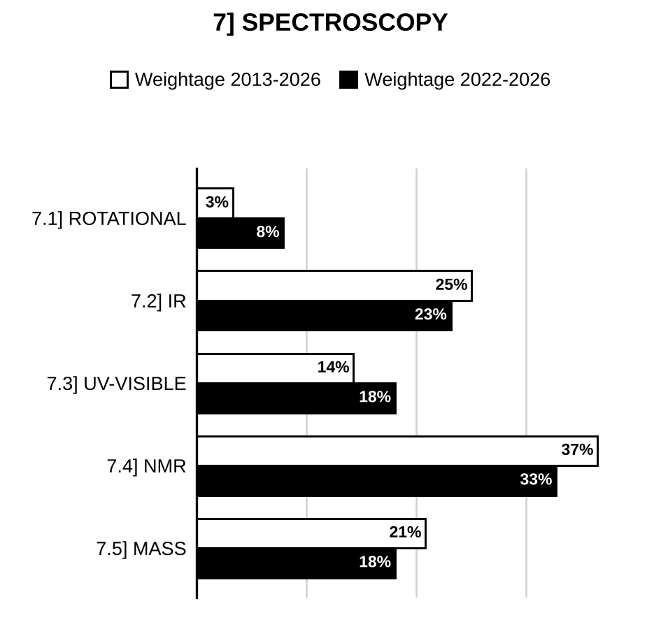
7.1] Rotational spectroscopy
8 questions
Calculate the rotational constant of a diatomic molecule if the moment of inertia is . (Given: Planck’s constant Js and velocity of light ) [2026/5m/8a(i)]
Which of the following molecules is/are active to rotational spectroscopy and why? , , , , [2025/5m/5d(i)]
The spacing between lines in the microwave spectrum of CO decreases by substituting C by C. Why? [2025/5m/5d(ii)]
Calculate the frequency of radiation required for a transition of J = 4 to J = 5 in the rotational spectrum of HCl. The rotation constant . [2025/5m/6b(i)]
Using the following data, calculate the bond length of HCl: ; 1 a.m.u. kg. [2024/5m/8c(iii)]
(A) , HCl, BrF, and Which of the above molecule(s) will show a microwave rotational spectrum? Justify your answer. (B) The first rotational absorption of was observed at while that of was observed at . Find their rotational constants. [2023/10m/8b(i)]
What is meant by rotational degrees of freedom for a molecule? Explain the selection rules for rotational spectra of molecules. [2007/10m/8c(i)]
(i) How will you account for the following mass spectral peaks in ? (m/z): 108, 107, 106, 105, 91, 79. Show the fragmentation pattern. (ii) The rotational spectrum of HCl molecule shows that the rotational lines are equally separated by . Calculate the internuclear bond length. (, ). [2003/20m/6c]
7.2] Vibrational (IR) spectroscopy
68 questions
How can the following pairs of compounds be distinguished based on the vibrational frequencies of their >C=O and >N–H bonds?
[Figure: Pair 1: ethyl benzoate (C6H5–CO–O–CH2–CH3) and phenyl propanoate (C6H5–O–CO–CH2–CH3). Pair 2: 4-bromo-N-methylbenzamide (4-Br–C6H4–CO–NH–CH3) and N-(4-bromophenyl)acetamide (4-Br–C6H4–NH–CO–CH3)] [2026/5m/5d(i)]
Arrange the following molecules in increasing order of vibrational frequency (): HF, HI, NO, CO. [2026/5m/8a(ii)]
Explain the specific vibrational frequencies (>C=O and –OH) in pentane-2,4-dione and 2-hydroxypropiophenone. [2026/5m/8a(iii)]
The fundamental vibrational frequency of HCl is . Calculate the fundamental vibrational frequency of DCl assuming the same bond strength. [2025/5m/6b(ii)]
The molecular formula of a compound is . The IR absorption frequencies are , and . Assign a structure for the compound. [2025/5m/6b(iii)]
An unknown organic compound with molecular formula displays a band at and a strong band at in the IR spectrum. The compound shows only two signals in 3:2 ratio in the H NMR spectrum. Find out the structure of the compound. Justify your answer. [2025/10m/8c(ii)]
Given below are the NMR spectral characteristics of two isomeric compounds with molecular formula : (1) H NMR: 2.0 (3H, s), 2.93 (2H, t), 4.3 (2H, t), 7.3 (5H, s) (2) H NMR: 1.23 (3H, t), 3.72 (2H, s), 4.13 (2H, q), 7.3 (5H, s) Both of these compounds exhibit a peculiar peak in IR spectra at . Deduce the structures of these two compounds. [2024/10m/7c(i)]
Ethanolic solution of compound I on irradiation leads to the formation of compounds II, III and IV. The resulting reaction mixture exhibits bands at 1787, 1740, 1715 and for . Assign these C=O stretching frequencies to the corresponding compounds giving reasons:
[Figure: Reaction: I (verbenone: 4,6,6-trimethylbicyclo[3.1.1]hept-3-en-2-one, α,β-unsaturated bicyclic ketone with the gem-dimethyl bridge) –hν, EtOH→ II (chrysanthenone: isomeric bicyclo[3.1.1] ketone with the C=O on the four-membered ring and the C=C(CH3) in the six-membered ring) + III (ethyl 3,7-dimethylocta-2,6-dienoate, (CH3)2C=CH–CH2–CH2–C(CH3)=CH–COOEt) + IV (ethyl 3,7-dimethylocta-3,6-dienoate, (CH3)2C=CH–CH2–CH=C(CH3)–CH2–COOEt)] [2024/15m/8a]
2-Chloro-2,3-dimethyl butane on dehydrohalogenation can lead to the formation of two products. Explain how the two can be distinguished using H NMR and IR spectral data. [2024/10m/8c(i)]
Compare the C=C stretching vibrations in the above compounds and give a suitable explanation for your answer.
[Figure: methylenecyclopropane (cyclopropane ring with exocyclic =CH2) and methylenecyclobutane (cyclobutane ring with exocyclic =CH2)] [2023/10m/5b]
The frequencies of vibration of the following molecules in their states are HCl: ; : ; DCl: and HD: . Calculate the energy change of the following reaction: Determine whether energy is liberated or absorbed. [Given: Veloci ]. [2023/10m/6a(i)]
The IR spectra of butyric acid and ethyl butyrate show sharp strong singlet absorption at and , respectively. By contrast, the IR spectrum of butyric anhydride shows a broad, sharp doublet at and . Why are these so different? [2023/5m/6a(ii)]
An organic compound having molecular formula gave following IR and NMR data: IR() = 1690; NMR(, 400 MHz): 1.11(t, J = 7Hz, 6H), 1.29(d, J = 7Hz 6H), 2.40 (g, J = 7Hz, 4H), 2.55(t, J = 7Hz, 2H), 2.65(t, J = 7 Hz, 2H), 3.12(septet, 1H), 7.21(d, J = 8Hz, 2H), 7.81(d, J = 8Hz, 2H). Determine the structure of the compound. [2023/10m/6c(i)]
How many fundamental vibrational frequencies would be expected to be observed in the IR spectrum of ? Sketch them. Identify the IR-active vibrations with justification. [2023/5m/8c(iii)]
(i) How will you distinguish between NH stretching absorption of a primary amine and a secondary amine by using IR spectroscopy? (ii) How will you distinguish among primary, secondary and tertiary alcohols on the basis of PMR spectroscopy? [2022/10m/5d]
Given below are the IR and NMR spectral characteristics of three compounds: (i) IR: ; NMR: 2.0 (s, 3H), 5.1 (s, 2H) and 7.3 (s, 5H) (ii) IR: ; NMR: 3.5 (s, 3H), 3.6 (s, 2H) and 7.4 (s, 5H) (iii) IR: 3200–2800 (various bands) and ; NMR: 2.75 (t, 2H), 2.95 (t, 2H), 7.4 (s, 5H) and 12.0 (s, 1H) Match each of these spectral data with one of the following structures:
[Figure: (1) methyl 4-methylbenzoate (para); (2) methyl phenylacetate ; (3) 4-ethylbenzoic acid, drawn as (para); (4) benzyl acetate ; (5) 3-phenylpropanoic acid ] [2022/10m/5e]
Among the following compounds and , which has more carbonyl stretching frequency in IR spectra? Explain:
[Figure: = 1-(cyclohex-1-en-1-yl)ethan-1-one (acetyl on an alkene carbon of cyclohexene); = 1-(2-methylcyclohex-1-en-1-yl)ethan-1-one (as with a CH3 on the other alkene carbon)] [2022/5m/8a(i)]
Complete the following reaction. Write the structures of , and , and explain how IR spectroscopy is helpful to distinguish among them:
[Figure: Reaction: 1,2-dimethylcyclohexan-1-ol (ring carbon bearing H3C and OH; adjacent ring carbon bearing CH3) –→ ] [2022/10m/8a(ii)]
The molecule obtained on treatment of acetone with dilute sodium hydroxide exhibits the above spectral data. Propose the structure of this molecule. IR: and NMR: 1.9 (s, 3H), 2.1 (s, 6H), 6.15 (s, 1H) [2021/15m/6a(i)]
Identify the compound in each of the above pairs, that can be expected to exhibit carbonyl stretching signal at higher frequency.
[Figure: Pair I: 5,6-dihydro-2H-pyran-2-one (δ-lactone with C=C conjugated to C=O: ring O–C(=O)–CH=CH–CH2–CH2) and 3,4-dihydro-2H-pyran-2-one (enol lactone: ring O–C(=O)–CH2–CH2–CH=CH–O); Pair II: and ] [2021/5m/6a(ii)]
A molecule with molecular formula exhibits only one signal at 1.2 ppm in NMR spectrum. This also exhibits a strong absorption at in IR spectrum. Propose structure for this molecule. [2021/10m/8b(i)]
Acetone exhibits only one carbonyl stretching frequency in IR spectrum whereas chloroacetone exhibits two at 1725 and . Explain why. [2021/5m/8c(ii)]
Arrange the following compounds in increasing order of out of plane C–H wagging vibrations, as shown, and give a suitable explanation to justify your answer:
[Figure: (A) 1,3-disubstituted benzene (R at C1 and C3) with only the isolated H at C2 drawn; (B) 1,2-disubstituted benzene (R at C1 and C2) with the four adjacent H drawn; (C) 1,3-disubstituted benzene (R at C1 and C3) with the three adjacent H (C4, C5, C6) drawn; (D) benzene with all six H drawn] [2020/10m/5d]
(i) Calculate C=O stretching frequency, dyne/cm.
(ii) A compound has molecular formula , showing absorption at 1380 . Identify the structure of the compound. [2020/15m/7a]
(i) What are metastable ions and how are they formed? The mass spectrum of toluene shows strong molecular ion peak at 91 and its fragment at 65. Calculate the value for metastable ion peak. (ii) Identify how vibrational spectroscopy might be used to monitor the progress of the following reaction:
[Figure: Reaction: 2,3-dimethylbut-1-ene () –conc. → 2,3-dimethylbut-2-ene ()] [2020/20m/7c]
How will you distinguish the two using IR-spectroscopy?
[Figure: I: 3,4-dimethylisoxazol-5(4H)-one (five-membered ring O1–N2=C3(CH3)–C4(H)(CH3)–C5(=O)–O1, the H on C4 drawn); II: 3,4-dimethylisoxazol-5(2H)-one (ring O1–N2(H)–C3(CH3)=C4(CH3)–C5(=O)–O1)] [2019/10m/5e]
On the basis of the given spectroscopic data, deduce the structure of : UV: nm (in ethanol) IR (): 3440 , 3360 , 3025 , 2920 , 1625 , 1380 , 810 H-NMR (ppm): , s, 3H , s, 2H , d, 2H ( Hz) , d, 2H ( Hz) [2019/15m/6a]
An organic compound with molecular formula exhibited the following spectroscopic data: IR (): 1830, 1760, 1120 H-NMR: , 2H, s , 2H, s Mass: = 148, 104, 76, 50 Deduce the structure of the compound and justify the formation of the above fragment ions. [2019/15m/7a]
How can you distinguish between the following pairs of compounds by using IR spectroscopy? (i) Salicylic acid and p-Hydroxy benzoic acid (ii) Propionaldehyde and Acetone. [2017/10m/5e]
How will you distinguish the following compounds by IR spectroscopy? Which will absorb more wave number in , and why?
[Figure: Two trans-decalin ketones drawn as fused chairs, C=O in the right ring with Cl on the adjacent ring carbon (3-chloro-trans-2-decalone): (I) Cl axial (pointing down), H equatorial; (II) Cl equatorial, H axial (pointing down)] [2017/5m/7a(iii)]
An unknown organic compound having molecular formula exhibited the following spectral data: UV: 270 nm; IR: H NMR: 7.6 (2H, d, J = 8 Hz), 6.9 (2H, d, J = 8 Hz), 3.9 (3H, s), 2.0 (3H, s). Deduce the structure of the compound. [2017/10m/8d]
How can cis- and trans-1,3-cyclohexane diol be distinguished using IR spectra? [2016/10m/6e]
Briefly explain why C = O bond stretch frequency occurs at for the carbonyl compound of an aliphatic ester and at for the carbonyl group of acid chloride. [2015/10m/5d]
An unknown compound, , did not dissolve in aqueous NaOH. The IR spectrum exhibited strong absorption at . The H NMR spectrum had signals at 7.2 (multiplet), 4.1 (quartet) and 1.3 (triplet). Elucidate the structure of the unknown compound. [2015/10m/6b]
One of the five compounds shown below was analysed by IR spectroscopy. Peaks were observed at (weak) and (strong). There was no signal between to . Which compound was analysed?
[Figure: Five compounds: 1 cyclohexanol; 2 cyclohexa-1,4-diene; 3 cyclohexanone; 4 cyclohex-3-en-1-one (C=O not conjugated with the ring C=C); 5 cyclohex-3-en-1-ol] [2015/10m/8e]
Account for the different carbonyl absorptions [ C = O] for the following compounds:
[Figure: acetophenone C6H5–CO–CH3, 1683 cm⁻¹; benzoyl bromide C6H5–CO–Br, 1812 cm⁻¹; ethyl benzoate C6H5–CO–OC2H5, 1720 cm⁻¹] [2014/10m/6a(ii)]
Assign structure to the compound with mol. formula , on the basis of the following spectral data: (i) UV (EtOH): 268, 264, 257, 243 nm (ii) IR: , , , (iii) H NMR: 7.1 – 7.3 (m, 5H), 5.0 (s, 2H), 1.96 (s, 3H) (iv) Mass: m/e ions: 150, 108, 91, 43. [2014/15m/7a]
An organic compound gives a strong absorption band at in IR spectrum. In proton NMR, it gives signal at 1.1 (triplet), 2.1 (singlet) and 2.3 (quartet). Write the likely structure of the compound and assign the values. [2013/20m/6a]
(i) Draw the rough-sketch of NMR spectra of . (ii) Which bond has higher frequency in IR spectra? (1) (2) [2012/12m/5c]
Which has higher stretching frequency in IR spectra? Explain. (i) OR (ii) [2012/15m/7b]
How will you differentiate the following pairs by IR spectra? (i) and (ii) and.
[Figure: (ii) second compound: oxirane (ethylene oxide), bridged by O] [2011/10m/5d]
When acetone is heated with NaOH (aq.) a product (A) is obtained which showed the following spectral features: IR (): 1695, 1620; H NMR (): 1.90 (S, 3H), 2.10 (S, 6H), 6.00 (S, 1H); Mass (m/e): 98, 83, 55 (100%), 53, 43, 39. Deduce the structure of A and interpret the spectral data. [2011/30m/8a]
How would you explain the carbonyl stretching frequencies given below the structure of the following compounds?
[Figure: cyclohexanone, 1727 ; cyclopentanone, 1754; cyclobutanone, 1802; cyclopropanone, 1825] [2011/15m/8c]
What is the range of fingerprint region in infrared spectrum. Write the range in which carbonyl group normally absorbs. [2010/10m/5f]
What characteristic bands will be observed in the ir spectrum of 1-phenylethanone? [2010/10m/8b]
Ethene and Ethyne unlike propene and propyne do not give C to C multiple bond stretching bonds in their IR spectrum. Explain. [2009/10m/6c(ii)]
The dehydration of 1, 2-dimethyl cyclohexanol (cis or trans) gives three alkenes. How can you distinguish them on the basis of their IR spectra? [2008/20m/5c(ii)]
Identify A-D in the following 2, 3-dimethyl-2-chlorobutane on E2 dehydrohalogenation gives how many products? Can they be distinguished by IR spectroscopy? Justify your answer. [2008/20m/6d]
How and are distinguished by IR spectroscopy and proton NMR spectroscopy? Give method for chemical separation of the two compounds. [2008/20m/8a]
(i) Grade the following transitions according to the amount of energy required and give explanation for your grading on the basis of Molecular Orbital Theory in a qualitative manner:- ; ; . (ii) The IR spectrum of p-nitrobenzyl alcohol, when recorded in the KBr phase, shows a medium, broad absorption at around 3200 . The same absorption shifts to 3450 as medium, sharp absorption when recorded in solution. Explain. [2007/20m/5c]
Using IR spectroscopy, indicate how will you differentiate between the following pairs of compounds? (i) Benzyl chloride and Benzyl Cyanide (ii) Aniline and N-methylaniline (iii) Cyclohexanone and Cyclohexanol (iv) Benzoic acid and Benzaldehyde (v) Styrene and Phenylacetylene. [2007/20m/7c]
The mass spectrum of a compound showed its at 177. Its IR spectrum showed peaks at 3000 (medium, sharp), 2700 (medium, sharp), 1660 (strong, sharp) and no other diagnostic absorptions. Its NMR spectrum showed signals at δ 1.25 (t, 6H), 3.4 (q, 4H), 6.6-7.4 (, 4H) and 9.7 (s, 1H). The compound analyzed for C 74.6%, H 8.6% [?] and N 8.1%. No evidence for the presence of S or halogens cound be found. Deduce the structure of this compound. [2007/15m/8c(iv)]
How many vibrational degrees of freedom will be observed for gaseous acetylene () and which of these will be infrared active? [2006/20m/5b]
How will you use IR and NMR spectroscopy to distinguish o- and p-hydroxyacetophenones? [2006/12m/6b]
How can NMR/IR be used to estimate enol content of the following? (1) (2) (3) [?].
[Figure: (3) bicyclo[2.2.1]heptane-2,3-dione (norbornane-2,3-dione, two adjacent C=O on the two-carbon bridge)] [2006/10m/8a(ii)]
Using the indicated spectral determination, how the following pairs of isomeric compounds can be differentiated? (i) and UV spectrum (ii) and IR spectrum (iii) and NMR spectrum (iv) [?] Any spectral method (v) and NMR spectrum.
[Figure: (iv) 4-(chloromethyl)benzonitrile (CN and para) and 3-chloro-5-methylbenzonitrile (CN at C-1, Cl at C-3, at C-5)] [2006/20m/8b]
Normal O – H stretch comes at about 3600 . What is the reduced mass for O – H? What happens to the reduced mass when atomic mass of each atom is doubled in turn; i.e., when O – H is converted to O – D and S – H? In fact both O – D and S – H stretch come at about 2500 . Why? [2005/20m/5a]
Indicate how IR spectrum can be used to distinguish:
[Figure: (i) 1-(cyclopent-1-en-1-yl)ethanone (acetyl on the ring double-bond carbon, conjugated) and 1-(cyclopent-3-en-1-yl)ethanone (acetyl on the sp3 ring carbon opposite the C=C, non-conjugated), each drawn with C(=O); (ii) dicyclohexylidenemethane (two cyclohexane rings joined by =C= , an allene) and bis(cyclohex-1-en-1-yl)methane (two cyclohexene rings joined through their double-bond carbons by )] [2005/20m/5b]
The natural product bullatenone was isolated in the 1950s and assigned the structure A. When compound A was synthesized, it was not identical with the natural sample. Predict the expected NMR spectrum of A. Given the full spectroscopic data available nowadays, but not in 1950s, say why A was wrong and suggest a better structure for bullatenone.
[Figure: A, “Alleged bullatenone”: 3-methyl-6-phenyl-2,3-dihydro-4H-pyran-4-one (six-membered ring: C-4 C=O, C-5=C-6 double bond with Ph on C-6, ring O, C-2 , C-3 bearing a )]
Spectra of bullatenone: Mass spectrum: m/z 188 (10%), Mol. formula: (), 105 (20%), 102 (100%), 77 (20%) IR spectrum: 1604 and 1705 NMR spectrum: 1.45(6H, s), 5.82(1H, s) 7.35(3H, m), 7.68(2H, m) [2005/15m/7a]
Indicate main stretch frequencies for functional groups present in acetyl acetone. What would happen to the spectrum if a small amount of organic base is added to it? [2005/20m/7b(ii)]
Using chemical and/or spectral methods (as indicated), show how the following can be differentiated: (i) and (Chemical methods only) (ii) (Chemical and spectral methods) (iii) (Spectral methods only)
[Figure: (ii) 2-phenylpropan-2-ol () and (2,4-dimethylphenyl)methanol (benzene ring with at C-1, at C-2 and at C-4); (iii) 2-hydroxyacetophenone (OH and ortho) and 4-hydroxyacetophenone (OH and para)] [2005/15m/8b]
(i) Infra-red spectrum of (2R. 3S) 2, 3-dichloro-pentane was recorded. Do you think that this spectra will be identical with the spectra of (2S. 3R) 2. 3-dichloro-pentane & (2R.3R)2, 3-dichioro-pentane? Give reasons for your answer. (ii) The mass spectrum of ethyl phenyl ether has a prominent peak at m/z 66. Describe mode of formation of the fragment responsible for this peak. [2004/20m/5c]
(i) Calculate the for the following using Wood ward-Fieser rule: (ii) Account for the changes in the carbonyl stretching frequency in the following.
[Figure: (i) (1) bicyclic α,β-unsaturated ketone: 4a-methyl-4,4a,5,6,7,8-hexahydronaphthalen-2(3H)-one skeleton with angular at C-4a, C=O at C-2, C=C between C-1 and C-8a, a on C-1 and an isopropyl group on C-7; (2) tricyclic tetraene: hydrophenanthrene skeleton with an angular at the junction of the left and middle rings, one C=C in the left ring (conjugated with the middle ring), two C=C in the middle ring, and one C=C in the right ring at the ring junction side, forming a conjugated system; (ii) three five-membered lactones: furan-2(5H)-one (C=C between C-3 and C-4, α,β-unsaturated) : 1750 ; γ-butyrolactone (saturated) 1770 ; furan-2(3H)-one (C=C between C-4 and C-5, next to the ring O) 1800 ] [2003/20m/6b]
Predict the position of absorption bands for the carbonyl group in the IR spectra of the following compounds:
[Figure: 1 = methyl benzoate (C6H5–CO–OCH3); 2 = 5,6-dihydro-2H-pyran-2-one (α,β-unsaturated six-membered lactone); 3 = benzoic acid; 4 = cyclohex-2-en-1-one; 5 = cyclopentanone; 6 = (E)-but-2-enal (crotonaldehyde, CH3–CH=CH–CHO)] [2002/15m/8a]
Deduce the structure of a compound on the basis of the spectral data given below: (i) Molecular formula: (ii) UV, : < 210 nm ( > 10,000), 280 nm ( = 900) (iii) IR, : 3300, 1710, 1670 (iv) NMR, : 2.21 (3H, s), 3.37 (2H, s), 3.76 (3H, s), 4.34 (2H, d), 6.80–7.18 (4H, dd) (One proton does not show, it exchanges with ) [2002/5m/8d]
Which compound A or B is expected to show lower stretching frequency for C = O group in the IR spectrum? (ii) How do trimeric phosphonitrillic halides compare with benzene in structure and reactivity?
[Figure: A = 4′-methoxyacetophenone (, para); B = 4′-nitroacetophenone (, para)] [2001/15m/8b(i)]
Spectral data of a compound (molecular formula ) are given below. Suggest a structure to this compound which is consistent to the spectral data. (i) UV; (EtOH; (): 256 nm (12 600), 324 nm (3,400) (ii) IR; (): 3175–3275 (broad). 1667 (strong), 1616, 1580, 1484, 1456, 1383, 1230, 1205, 1151, 1115 and 1029 (iii) NMR; (): 10.83 (1H, S) which disappears on addition of 9.59 (1H, S) 7.31–6.79 (4H, m) (iv) Ms; m/z (Relative abundance): 122 ( 100%), 121 (96%) 93 (18.5%), 29 (31%) [2001/15m/8d]
(i) How could you use IR spectroscopy to tell if preparation of 5-cholesten-3-one from cholesterol by Jones oxidation is complete?
[Figure: Partial steroid skeleton (rings A and B with angular CH3 at C10, C5=C6 double bond, rest of the molecule shown by a wavy bracket): 3-OH compound (cholesterol) –(Jones Oxid.)→ the corresponding 3-ketone (5-cholesten-3-one, C3=O)]
(ii) Distinguish between the following pairs by IR. (A) and (B) and [2000/15m/8d]

Preparing for Prelims too?
Our UPSC Prelims Topic-wise PYQ eBook covers every Prelims question with detailed explanations, so you can see exactly where UPSC asks from.
View the BookDownload Free Sample7.3] Electronic (UV-visible) spectroscopy
38 questions
Using the Woodward-Fieser rules, calculate the values of the following compounds:
[Figure: Two tricyclic compounds on a perhydrophenalene skeleton (upper-left, upper-right and lower six-membered rings all sharing one central carbon, C9b). First (triene): C=C in the upper-left ring between the carbon bearing CH3 and the carbon shared by the upper-left and lower rings; the central carbon is double-bonded to the carbon shared by the upper-right and lower rings; that carbon is bonded to a lower-ring carbon that is double-bonded to the next lower-ring carbon (linear conjugated triene C=C–C=C–C=C, three C=C in all); remaining carbons sp3. Second (dienone): the same CH3-bearing C=C in the upper-left ring and the same central-carbon C=C to the carbon shared by the upper-right and lower rings, which is bonded to a C=O carbon in the upper-right ring (C=C–C=C–C=O); lower ring saturated] [2026/5m/5d(ii)]
Explain the effect of conjugation on and intensity due to n- and – transitions in 3-buten-2-one compared to acetone. [2026/5m/8c(i)]
Explain the formation of charge-transfer complex between (A) picric acid and anthracene, and (B) tetracyanoethylene and aniline, using electronic spectroscopy. [2026/5m/8c(ii)]
Acetone shows a weak absorption at 280 nm and a strong absorption at 190 nm in the UV spectrum. Account for the observation. [2025/5m/7a(i)]
Using Woodward-Fieser rules, calculate for the following compounds:
[Figure: A: 3-methylpent-3-en-2-one, CH3–CH=C(CH3)–CO–CH3. B: 2-cyclohexylidenecyclohexan-1-one (cyclohexanone with an exocyclic C=C at C2 to a second cyclohexane ring). C: 1-acetylcyclopentene (1-(cyclopent-1-en-1-yl)ethan-1-one). D: fused bicyclic dienone (hexahydronaphthalenone): a cyclohexenone ring with C=O, its α,β-C=C running to a ring-fusion carbon (exocyclic to the second ring), and a methyl on the carbon adjacent to the other (sp3) ring-fusion carbon; the second ring carries one more C=C conjugated with the enone C=C (C=C–C=C–C=O system, the extra C=C in the other ring)] [2025/10m/7a(ii)]
In UV spectra of the following pairs, which compound will have higher ?
[Figure: (i) A: 4′-methylacetophenone (COCH3 and CH3 para); B: 2′-methylacetophenone (COCH3 and CH3 ortho). (ii) A: 1,1′-bi(cyclohex-1-enyl) (two cyclohexenes joined at C1, conjugated diene with each C=C endocyclic); B: 1,2-dicyclohexylideneethane (cyclohexylidene=CH–CH=cyclohexylidene, both C=C exocyclic). (iii) A: (Z)-penta-1,3-diene, CH2=CH–CH=CH–CH3 with CH3 and the vinyl group cis; B: (E)-penta-1,3-diene (CH3 trans to the vinyl group). (iv) A: 4-nitroaniline; B: 3-nitroaniline. (v) Two isomeric bicyclic (decalin-type) enones, each with an isopropenyl group (C(CH3)=CH2) on the saturated ring. A: ketone ring = ring-fusion CH – CH(CH3) – C(=O) – CH=C(CH3) – ring-fusion CH, i.e. endocyclic α,β-enone with CH3 on the β-carbon and CH3 on the α′-carbon; both ring-fusion carbons sp3. B: ketone ring = ring-fusion C=C(CH3) – C(=O) – CH2 – CH(CH3) – ring-fusion CH, i.e. tetrasubstituted α,β-C=C whose β-carbon is a ring-fusion carbon (C=C exocyclic to the other ring), CH3 on the α-carbon and on the carbon next to the other ring-fusion carbon] [2024/10m/5d]
Calculate the value of in the following compounds using Woodward-Fieser rules:
[Figure: Three tricyclic ketones on the perhydrophenanthrene skeleton (angularly fused rings: left, middle, right). A: C=O in the right ring on the carbon next to the lower middle/right ring-fusion carbon; that fusion carbon is double-bonded to the adjacent lower middle/left ring-fusion carbon (α,β-C=C), which is bonded to the other middle/left ring-fusion carbon, itself double-bonded to the next middle-ring carbon (CH); both C=C lie in the middle ring (C=C–C=C–C=O); the remaining middle-ring carbons are CH2 and the upper middle/right fusion CH (sp3). B: C=O in the right ring next to the lower middle/right fusion carbon; α,β-C=C is the middle/right ring-fusion bond, extended by a C=C between the next two middle-ring carbons (CH=CH); the middle/left fusion carbons are sp3 CH. C: C=O in the middle ring, bonded to a CH2 (towards the upper right ring, which is saturated and fused at two sp3 CH) and to a left/middle ring-fusion carbon that is double-bonded into the left ring; the left ring carries a second conjugated C=C (homoannular diene C=C–C=C conjugated with C=O), its other ring-fusion carbon sp3 CH and one CH2] [2024/15m/7a]
Draw the structural formula of a ketone with MF that absorbs in the UV with nm. [2023/5m/8a(ii)]
Calculate the values of the following compounds:
[Figure: (1) a steroid with four conjugated C=C bonds: C3=C4 in ring A, C5=C6 and C7=C8 in ring B, and C9=C11 in ring C (cholesta-3,5,7,9(11)-tetraene skeleton), angular CH3 at C-13 and side chain at C-17, no methyl drawn at C-10; (2) a bicyclic decalin-type triene: 1-vinyl-2,3,4,6,7,8-hexahydronaphthalene – the vinyl group CH=CH2 on C-1, C1=C8a double bond (at the ring fusion carbon) and C4a=C5 double bond in the other ring, so that CH2=CH–C1=C8a–C4a=C5 is one conjugated chain] [2022/10m/8b(i)]
Explain why 1,3-butadiene exhibits a lower for transitions compared to that of 1,3,5-hexatriene. [2022/5m/8b(ii)]
(1) A compound with MF exhibits two doublets ( Hz) in its PMR spectrum. Suggest a suitable structure along with other possible structures. (2) How can the structures of and be decided based on their UV spectral data? [ nm () and nm ()].
[Figure: = β-ionone: 2,6,6-trimethylcyclohex-1-ene ring bearing at C-1 a –CH=CH–CO–CH3 (E) chain; = (E)-4-(cyclohex-1-en-1-yl)but-3-en-2-one: cyclohexene bearing –CH=CH–CO–CH3 on an alkene carbon] [2022/10m/8c(ii)]
The UV spectrum of acetone exhibits two signals of different intensities, one at 280 nm and the other at 190 nm. Assign corresponding electronic transitions to the observed signals. [2021/5m/8b(ii)]
(i) Dichloromethane has a chemical shift of 532 Hz from TMS (tetramethylsilane) on a 100 MHz spectrometer. What will be the chemical shift in ppm? What would the chemical shift be in ppm and Hz on a 600 MHz spectrometer? (ii) 3-Buten-2-one gives two peaks in its ultraviolet spectrum at 213 nm (ε = 7100) and 320 nm (ε = 27). What type of transition is responsible for each absorbance? Account for the difference in molar absorptivity for the two absorptions. [2020/15m/8a]
Predict for the following molecules: (i) (ii)
[Figure: (i) 6-methoxy-1-tetralone (6-methoxy-3,4-dihydronaphthalen-1(2H)-one: on the aromatic ring, para to the ring-fusion carbon bearing the C=O); (ii) 1-(cyclohex-2-en-1-ylidene)propan-2-one (cyclohexene ring with C2=C3 in the ring, exocyclic C1=CH–C(=O)–CH3)] [2019/10m/5d]
How do the UV spectral data 296 nm, 10700 and 281 nm, 20800 help in deciding between structures and ?
[Figure: X: β-ionone (4-(2,6,6-trimethylcyclohex-1-en-1-yl)but-3-en-2-one: ring C=C conjugated with side-chain trans CH=CH–C(=O)CH3; gem-dimethyl at C6 and CH3 at C2 of the ring); Y: 4-(cyclohex-1-en-1-yl)but-3-en-2-one (cyclohexene ring, no methyls, ring C1=C2 with C1 bearing the trans chain)] [2018/10m/8d]
Explain charge-transfer spectra by taking a suitable donor-acceptor type complex. [2018/10m/8e]
How can you distinguish between cis and trans isomers of stilbenes from UV spectral study? Explain. [2017/10m/7c]
Which of the following structures has 229 nm? Give the calculations of each:
[Figure: (i) cyclohexa-1,3-diene ring with CH3 on a diene terminus carbon (top) and isopropyl on the sp3 carbon para to it (bottom); double bonds on the upper-left edge (from the CH3-bearing carbon) and on the right vertical edge (α-phellandrene type, homoannular diene); (ii) cyclohexene ring with an exocyclic =CH2 at the top carbon, a ring C=C on the right vertical edge conjugated with it, and isopropyl at the bottom carbon (β-phellandrene type, heteroannular/exocyclic diene); (iii) myrcene (7-methyl-3-methylene-octa-1,6-diene: CH2=CH–C(=CH2)–CH2–CH2–CH=C(CH3)2); (iv) 2-cyclohexylidenecyclohexanone (C=O conjugated with the exocyclic C=C to a second cyclohexane ring); (v) bicyclic cross-conjugated dienone: decalin-type skeleton with C=O in the left ring, a C=C between the carbonyl α-carbon and the ring-fusion carbon (bottom) and a second C=C on the other side of the carbonyl (upper left); angular CH3 on the upper ring-fusion carbon; CH3 groups on the two adjacent carbons of the right ring] [2016/15m/8a]
Calculate for the following compounds:
[Figure: (1) bicyclic (decalin-skeleton) heteroannular diene: one C=C in the left ring and a second C=C from the ring-fusion carbon into the right ring, conjugated with the first (transoid), with a CH3 on the left-ring alkene carbon adjacent to the ring fusion; (2) bicyclic homoannular diene: both C=C in the left ring, one of them ending at the ring-fusion carbon (C4=C4a) and the other at C2=C3, with an angular CH3 on the other ring-fusion carbon (C-8a)] [2014/5m/8g]
Calculate the for the following compounds using Woodward – Fieser rule:
[Figure: (i) 6-acetoxy-6-methylcyclohexa-2,4-dien-1-one (ring C=O at C1; C6 sp3 bearing and ; ring double bonds C2=C3 and C4=C5); (ii) bicyclic enone: cyclopentenone fused to a cyclohexane — five-membered ring with C=O, the carbon next to it bearing and attached to a ring-fusion CH, the other side of the C=O a ring CH= double-bonded to the other ring-fusion carbon (1-methyl-1,4,5,6,7,7a-hexahydro-2H-inden-2-one); (iii) 3,4,5-trihydroxybenzoic acid (gallic acid); (iv) 1-tetralone (3,4-dihydronaphthalen-1(2H)-one)] [2011/15m/8b]
What changes do you expect in the electronic spectrum of acetoacetic ester in hexane and ether. [2010/10m/5d]
An organic compound with molecular formula gives following spectral characteristics.
| Electronic spectrum | at 235 nm |
| Infrared spectrum | 3035, 3000, 2925, 1640, 990, 915, 740 and 615 |
| NMR spectrum | at 3.1(2H), 4.8(1H), 5.1(1H), 5.8(1H), 7.1(5H) |
| Mass spectrum | m/e at 117 |
Suggest a possible structure with probable justification for each spectral feature. [2010/30m/8a]
ion shifts transition of to higher energy. Explain. [2009/12m/6b(i)]
Polar solvent shifts of transition of to higher value. Explain. [2009/12m/6c(i)]
Using Woodward-Feiser rule predict the for the following in their electronic spectra:
[Figure: (A) six-membered ring with at C-1 and C-4 (opposite corners) and a ring C=C at the lower right; the remaining ring bonds are obscured by the watermark (apparently 1,4-dimethylcyclohexa-1,3-diene [?]); (B) bicyclic hexahydronaphthalene with a C=C at the ring fusion (C-8a=C-1), C-1 bearing a vinyl group printed “CH = H2” (i.e. [?]), and a second C=C in the other ring adjacent to the ring fusion] [2008/20m/5a(i)]
The geometric isomers of stilbene show at 278 and 294 nm. Assign each value to appropriate isomer and sketch them. [2008/20m/5d(i)]
(i) Grade the following transitions according to the amount of energy required and give explanation for your grading on the basis of Molecular Orbital Theory in a qualitative manner:- ; ; . (ii) The IR spectrum of p-nitrobenzyl alcohol, when recorded in the KBr phase, shows a medium, broad absorption at around 3200 . The same absorption shifts to 3450 as medium, sharp absorption when recorded in solution. Explain. [2007/20m/5c]
Predict which of the following pairs of compounds will have higher values and why? (i) and (ii) [?] and aniline (iii) Trans and (iv) Piperidine and [?] (v) [?] and [?].
[Figure: (ii) a cyclohexane ring bearing a substituent drawn as “—O” (label garbled; apparently cyclohexylamine [?]); (iv) 1-(cyclohex-1-en-1-yl)piperidine (the enamine: piperidine N bonded to the double-bond carbon of a cyclohexene ring); (v) cyclohexa-1,4-diene and cyclohexa-1,3-diene] [2007/20m/7b]
How do you assay bulk drugs using UV-visible spectroscopy? [2007/10m/8c(ii)]
Illustrate Woodward–Fieser Rules to calculate the expected of enones in the uv-spectrum. [2005/10m/7c]
An α, β-unsaturated ketone of relative molecular mass 110 has an absorption in U.V at 215 (). A solution of this ketone showed absorbance A = 2.0 with 1 cm cell. Calculate concentration of keton in the solution expressed in g/l. [2004/20m/5a]
U.V visible spectrum of an α, β-unsaturated carbonyl compound was recorded first in cyclohexane as solvent. What will be the effect on if the spectra of the same compound is taken in ethanol as solvent? Give reasons for your answer. [2004/20m/5b]
(i) Calculate the for the following using Wood ward-Fieser rule: (ii) Account for the changes in the carbonyl stretching frequency in the following.
[Figure: (i) (1) bicyclic α,β-unsaturated ketone: 4a-methyl-4,4a,5,6,7,8-hexahydronaphthalen-2(3H)-one skeleton with angular at C-4a, C=O at C-2, C=C between C-1 and C-8a, a on C-1 and an isopropyl group on C-7; (2) tricyclic tetraene: hydrophenanthrene skeleton with an angular at the junction of the left and middle rings, one C=C in the left ring (conjugated with the middle ring), two C=C in the middle ring, and one C=C in the right ring at the ring junction side, forming a conjugated system; (ii) three five-membered lactones: furan-2(5H)-one (C=C between C-3 and C-4, α,β-unsaturated) : 1750 ; γ-butyrolactone (saturated) 1770 ; furan-2(3H)-one (C=C between C-4 and C-5, next to the ring O) 1800 ] [2003/20m/6b]
p-nitroaniline exhibits a pronounced red shift in the K-band, which is not observed in o or m-isomer. Explain. [2002/5m/8b(i)]
Calculate the for the triene (A) which contains both a heteroannular and homoannular diene. Your calculation should be based on the homoannular diene.
[Figure: A = steroid triene (cholestane skeleton, at C17, angular methyls): two conjugated C=C in ring A and one C=C in ring B (C6=C7) conjugated with them, so that ring A holds a homoannular diene and rings A/B a heteroannular diene [?]] [2002/15m/8b(ii)]
When acetone is treated with a base, a higher boiling liquid (b.p. 130° C) can be isolated from the reaction mixture. The spectral data of this compound are as follows: IR, (): 1620, 1695; UV; max: 238 nm ( 11,700); 1H NMR; : 1.9 (3H, S), 2.1 (6H, S), 6.15 (1H, S); MS; m/z (R.A.): 98 (49%), 83 (90%); 55 (100%); 43 (78%). Deduce the structure of the compound. [2001/20m/5d]
The following dienes in ethanol have ’s at 231 nm ( 21,000), 236 nm ( 12,000). 245 nm ( 18,000), 265 nm ( 6,400) and 282 nm ( 11,900), Indicate which diene corresponds to which spectrum.
[Figure: Five dienes: (1) steroid (angular methyls, R at C17) with a heteroannular diene (C7=C8 and C9=C11); (2) steroid (angular methyls, R at C17) with a homoannular diene in ring B; (3) cyclohexadiene ring with CH3 at C1 and at C4 (p-menthadiene, α-terpinene type [?]); (4) cyclohexadiene ring with CH3 at C1 and CH3 [?] at C4 (1,4-dimethylcyclohexa-1,3-diene type [?]); (5) 1,1′-bi(cyclohex-1-enyl) (two cyclohexene rings joined through their C1 atoms)] [2001/15m/8a]
(i) Phenol is a weak acid with pKa = 10.0. It has = 210 nm in ethanol. When dil NaOH is added to the solution, the absorption increases to = 235 nm. How do you account for this shift. (ii) Calculate for the following:
[Figure: (A) 6,7,8,8a-tetrahydronaphthalen-2(1H)-one: bicyclic dienone, ketone C=O at C2 of the left ring, C3=C4 double bond in the left ring conjugated with a C4a=C5 double bond exocyclic to the left ring (in the right ring); (B) 2,4-dihydroxy-3,5-dimethylacetophenone: benzene ring with C(=O)CH3 at C1, OH at C2, Me at C3, OH (printed HO) at C4, Me at C5, H at C6] [2000/15m/8c]
7.4] NMR spectroscopy
85 questions
Explain and compare the chemical shifts of hydrogens in H NMR spectra of ethene and ethyne. [2026/5m/5e(i)]
Discuss the spin-spin interactions and coupling constants in H NMR spectrum of furfuraldehyde. [2026/5m/8b(i)]
Distinguish between (A) anthracene and phenanthrene, and (B) pure ethanol and ethanol in presence of trace amount of acidic impurity, based on H NMR spectra. [2026/5m/8b(ii)]
How can you differentiate between 2-nitroacetophenone and 3-nitroacetophenone based on approximate chemical shifts and spin-spin interactions? (Consider only ortho-coupling) [2026/5m/8b(iii)]
In a 100 MHz NMR instrument, a particular set of protons absorbs at with Hz. Find the chemical shift (in Hz) and the coupling constant J in a 500 MHz instrument for the same set of protons. [2025/5m/5e(i)]
Estimate the expected splitting (coupling constant J in Hz) for the lettered protons in the H NMR spectrum of the following compounds: (ii) Compare the chemical shifts of the labelled protons and in the H NMR spectrum of the following compounds and justify your answer:
[Figure: (i) (1) A: 1-bromo-1-chloroethene, (H^a on the same side as Br, H^b on the same side as Cl). (2) B: trans-1,2-dichloroethene, H^a(Cl)C=C(Cl)H^b with H^a and H^b trans. (3) C: (propargyl chloride)]
[Figure: (ii) D: methyl vinyl ether CH2=CH–OCH3 with H^a the terminal =CH2 proton cis to OCH3. E: methyl vinyl ketone CH2=CH–COCH3 with H^b the terminal =CH2 proton cis to COCH3] [2025/10m/8a(i)]
Count the number of peaks observed in the H NMR spectrum of the following compounds. Justify your answer:
[Figure: (1) 2-chlorobutane, CH3–CH(Cl)–CH2CH3 with Cl on a wedge. (2) 1,6-dimethylcyclohexa-1,4-diene: Me on sp2 C1 and Me on a wedge at the adjacent sp3 C6; C=C at C1=C2 and C4=C5; C3 is CH2] [2025/5m/8a(iii)]
A halogenated ester shows peak at m/z 166 (10%) and M+2 peak at m/z 168 (9.8%) in mass spectrum. H NMR spectrum of this compound shows two triplets and a singlet at 2.9, 3.6 and 3.8 ppm, respectively in the intensity ratio 1:1:1.5. Deduce the structure of the compound. Justify your answer. [2025/10m/8b(i)]
An unknown organic compound with molecular formula displays a band at and a strong band at in the IR spectrum. The compound shows only two signals in 3:2 ratio in the H NMR spectrum. Find out the structure of the compound. Justify your answer. [2025/10m/8c(ii)]
In H NMR spectrum of the following compounds, how many signals will be observed? In each case, label and arrange the hydrogens in the order of increasing chemical shift.
[Figure: I: anthracene; II: cyclohexa-1,3-diene; III: norbornadiene (bicyclo[2.2.1]hepta-2,5-diene)] [2024/10m/5e]
Given below are the NMR spectral characteristics of two isomeric compounds with molecular formula : (1) H NMR: 2.0 (3H, s), 2.93 (2H, t), 4.3 (2H, t), 7.3 (5H, s) (2) H NMR: 1.23 (3H, t), 3.72 (2H, s), 4.13 (2H, q), 7.3 (5H, s) Both of these compounds exhibit a peculiar peak in IR spectra at . Deduce the structures of these two compounds. [2024/10m/7c(i)]
2,2-Dimethyl cyclopropanone undergoes ring opening when attacked by methoxide ion and the product obtained possesses the following spectral data: IR (, ): 1740, 1160 H NMR (): 3.6 (3H, s), 1.2 (9H, s) Mass (m/z): 116, 85, 59, 31 Deduce the structure of the product with reasons. Write down the structure of another possible product. [2024/10m/8b(i)]
Arrange the following compounds in the order of increasing coupling constant values ():
[Figure: Three methyl but-2-enoate / methacrylate-type alkenes: I: C(Ha)(CH3)=C(COOCH3)(Hb) with Ha cis to COOCH3 and trans to Hb (Ha top-left, CH3 bottom-left, COOCH3 top-right, Hb bottom-right). II: C(Ha)(Hb)=C(COOCH3)(CH3), geminal Ha/Hb (Ha top-left, Hb bottom-left, COOCH3 top-right, CH3 bottom-right). III: C(CH3)(Hb)=C(COOCH3)(Ha) (CH3 top-left, Hb bottom-left, COOCH3 top-right, Ha bottom-right), Ha and Hb cis] [2024/5m/8b(ii)]
2-Chloro-2,3-dimethyl butane on dehydrohalogenation can lead to the formation of two products. Explain how the two can be distinguished using H NMR and IR spectral data. [2024/10m/8c(i)]
How many signals would you expect in the NMR spectrum of above compounds? Mark these protons.
[Figure: (A) 1-[4-(allyloxy)phenyl]ethan-1-one: benzene ring bearing CH3C(=O)– at one position and –O–CH2–CH=CH2 at the para position; (B) 1,2-dimethylcyclopropane drawn as a triangle with Me on the top carbon (pointing up) and Me on the lower-right carbon (pointing down); no wedges shown] [2023/10m/5a]
An organic compound having molecular formula gave following IR and NMR data: IR() = 1690; NMR(, 400 MHz): 1.11(t, J = 7Hz, 6H), 1.29(d, J = 7Hz 6H), 2.40 (g, J = 7Hz, 4H), 2.55(t, J = 7Hz, 2H), 2.65(t, J = 7 Hz, 2H), 3.12(septet, 1H), 7.21(d, J = 8Hz, 2H), 7.81(d, J = 8Hz, 2H). Determine the structure of the compound. [2023/10m/6c(i)]
Assign and arrange the lettered protons in the increasing order of their chemical shift value in NMR spectrum.
[Figure: 4,5-dihydrooxepine ring: O1; C2 bearing Ha, double-bonded to C3 bearing Hb; C4 (sp3) bearing a CHO group whose aldehyde H is Hc; C5 is CH2; C6 bearing an acetyl group C(=O)CH3 whose methyl is labelled CH3d (Hd), double-bonded to C7 bearing He; C7 bonded back to O1] [2023/10m/6c(ii)]
Estimate the expected splitting (J in Hz) for the lettered protons in the above compounds:
[Figure: (A) 1,1-dichloroethene, (Ha and Hb geminal on one carbon); (B) 1,1-dichloro-2-methylenecyclopentane: cyclopentane with exocyclic =CHaHb on one ring carbon and two Cl on the adjacent ring carbon; (C) 3-methoxycyclohexene: OCH3 on the sp3 ring carbon C-3, Ha on the adjacent alkene carbon C-2 and Hb on the other alkene carbon C-1] [2023/5m/8b(ii)]
In the NMR spectrum of acetyl acetone the height of the integration curve at methylene () signal was found to be 10 mm and that of methine (= CH) signal was 22 mm. Calculate the percentage of keto and enol form in the sample. [2023/10m/8c(i)]
(i) How will you distinguish between NH stretching absorption of a primary amine and a secondary amine by using IR spectroscopy? (ii) How will you distinguish among primary, secondary and tertiary alcohols on the basis of PMR spectroscopy? [2022/10m/5d]
Given below are the IR and NMR spectral characteristics of three compounds: (i) IR: ; NMR: 2.0 (s, 3H), 5.1 (s, 2H) and 7.3 (s, 5H) (ii) IR: ; NMR: 3.5 (s, 3H), 3.6 (s, 2H) and 7.4 (s, 5H) (iii) IR: 3200–2800 (various bands) and ; NMR: 2.75 (t, 2H), 2.95 (t, 2H), 7.4 (s, 5H) and 12.0 (s, 1H) Match each of these spectral data with one of the following structures:
[Figure: (1) methyl 4-methylbenzoate (para); (2) methyl phenylacetate ; (3) 4-ethylbenzoic acid, drawn as (para); (4) benzyl acetate ; (5) 3-phenylpropanoic acid ] [2022/10m/5e]
A compound () containing C, H and O has molecular weight 102 and displays two signals in the NMR spectrum at 1.1 (d) and 3.55 (septet) in the integral ratio of 6 : 1. Treatment of with 1 mole of HI gives rise to and . In the IR spectrum, gives a strong absorption band at , whereas its NMR spectrum shows signals at 1.05 (d, 6H), 3.6 (septet, 1H) and 4.4 (s, 1H, disappeared with ). The NMR spectrum of gives signals at 1.9 (d, 6H) and 4.25 (septet, 1H). Reaction of with excess of HI gives only . Identify , and . Write all the reactions involved. [2022/10m/7c(i)]
Three isomeric compounds having MF give positive 2,4-DNP test and display the following NMR spectral characteristics. Identify the compounds. Among them, one isomeric compound on treatment with KOH (concentrated) gives two products. Write their structures also: (1) A triplet at 1.05 and a quartet at 2.47 (2) Two singlets (3) A doublet at 1.0, a singlet at 2.1 and a septet at 2.2. [2022/10m/7c(ii)]
(1) A compound with MF exhibits two doublets ( Hz) in its PMR spectrum. Suggest a suitable structure along with other possible structures. (2) How can the structures of and be decided based on their UV spectral data? [ nm () and nm ()].
[Figure: = β-ionone: 2,6,6-trimethylcyclohex-1-ene ring bearing at C-1 a –CH=CH–CO–CH3 (E) chain; = (E)-4-(cyclohex-1-en-1-yl)but-3-en-2-one: cyclohexene bearing –CH=CH–CO–CH3 on an alkene carbon] [2022/10m/8c(ii)]
2,4-Pentadione exhibits five signal in NMR spectrum. Account for the observation. Also write the approximate chemical shift. [2021/10m/5a]
The molecule obtained on treatment of acetone with dilute sodium hydroxide exhibits the above spectral data. Propose the structure of this molecule. IR: and NMR: 1.9 (s, 3H), 2.1 (s, 6H), 6.15 (s, 1H) [2021/15m/6a(i)]
A molecule with molecular formula exhibits a broad band at in IR spectrum. Its mass spectrum exhibits base peak at m/z 135 and the NMR spectrum exhibits the following signals: 1.3 (d, 6H); 2.4 (s, 3H), 3.4 (m, 1H), 4.6 (s, exchangeable), 6.6 (s, 1H), 6.8 (d, 1H) and 7.1 (d, 1H). Deduce the structure. [2021/15m/7c]
A molecule with molecular formula exhibits only one signal at 1.2 ppm in NMR spectrum. This also exhibits a strong absorption at in IR spectrum. Propose structure for this molecule. [2021/10m/8b(i)]
Predict the number of signals, nature of the signals (s/d/t/m) and approximate chemical shifts in NMR spectrum of methyl propenoate. [2021/10m/8c(i)]
Consider the two methyl groups labelled as Ⓐ and Ⓑ in the following compound, and answer the given questions: (i) Which methyl group would exhibit a higher chemical shift in H NMR spectrum? (ii) Offer a suitable explanation to justify your choice of methyl group. (iii) Predict the approximate chemical shift values for both the methyl groups.
[Figure: 7,7-dimethylbicyclo[2.2.1]hept-2-ene (7,7-dimethylnorbornene) drawn with the C7 bridge at the top; methyl Ⓑ drawn to the left (over the saturated side) and methyl Ⓐ drawn to the right (over the C=C side, which is on the right of the drawing)] [2020/10m/5e]
(i) Dichloromethane has a chemical shift of 532 Hz from TMS (tetramethylsilane) on a 100 MHz spectrometer. What will be the chemical shift in ppm? What would the chemical shift be in ppm and Hz on a 600 MHz spectrometer? (ii) 3-Buten-2-one gives two peaks in its ultraviolet spectrum at 213 nm (ε = 7100) and 320 nm (ε = 27). What type of transition is responsible for each absorbance? Account for the difference in molar absorptivity for the two absorptions. [2020/15m/8a]
An organic compound X was erroneously assigned the following structure:
[Figure: Structure X: pyran-4-one ring: C4=O at top, C3 (next to C=O, on the right) bearing a CH3, C2 = CH2 bonded to ring O1, C6 (bonded to O1, lower left) bearing Ph and double-bonded to C5 (C5=C6, left edge), i.e. 3-methyl-6-phenyl-2,3-dihydro-4H-pyran-4-one]
The spectral data of the organic compound are given below. On the basis of the spectral data, justify why X is definitely a wrong structure and write the correct structure compatible with the spectral data: IR (): 1705, 1604 H NMR: (ppm) 1.43 (s, 6H), 5.82 (s, 1H), 7.35 (m, 3H), 7.68 (m, 2H) MS: 188 (10%) (high resolution confirms ), 105 (20%), 102 (100%), 77 (20%) [2020/20m/8c]
On the basis of the given spectroscopic data, deduce the structure of : UV: nm (in ethanol) IR (): 3440 , 3360 , 3025 , 2920 , 1625 , 1380 , 810 H-NMR (ppm): , s, 3H , s, 2H , d, 2H ( Hz) , d, 2H ( Hz) [2019/15m/6a]
An organic compound having molecular formula exhibited the following H-NMR data (ppm): , s , d ( Hz) , d ( Hz) Deduce the structure of the compound. [2019/15m/8a]
How will you distinguish various isomers of bromochloroethylene on the basis of H-NMR spectroscopy? [2019/15m/8b]
An organic compound (MF ) exhibits the following spectral characteristics: UV (: 280 nm) IR : 1715 H NMR (δ, ppm): 0.90 (3H, t), 1.60 (2H, m), 2.20 (3H, s), 2.40 (2H, t) Mass (): 86, 71, 58, 43 (100%) Deduce the structure of the compound and explain the principal ions observed in the mass spectrum. [2018/15m/4d]
A signal has been reported to occur at 120 Hz downfield from TMS in an NMR spectrometer with a 60 MHz operating frequency. (i) What is its chemical shift? (ii) What would its chemical shift be in an instrument operating at 100 MHz? (iii) How many hertz downfield from TMS would the signal be in a 100 MHz spectrometer? [2018/10m/5d]
How many H NMR signals do you expect for 1-bromopropane and 2-bromopropane? Give their splitting pattern. [2017/5m/7a(ii)]
An unknown organic compound having molecular formula exhibited the following spectral data: UV: 270 nm; IR: H NMR: 7.6 (2H, d, J = 8 Hz), 6.9 (2H, d, J = 8 Hz), 3.9 (3H, s), 2.0 (3H, s). Deduce the structure of the compound. [2017/10m/8d]
Assign the observed chemical shift values of 1.22, 1.88, 4.13, 5.81 and 6.95 for the following compound:
[Figure: ethyl (E)-but-2-enoate (ethyl crotonate): H3C and COOCH2CH3 on opposite carbons of C=C, trans to each other; each alkene carbon also bears H] [2016/10m/7d]
Draw structures, label equivalent protons and indicate the number of signals in each of the following: (i) Mesitylene (ii) p-ethylbenzene (iii) Isopropylbenzene. [2016/10m/8c]
Distinguish between the following compounds using proton NMR spectra: (i) 1-bromopropane and 2-bromopropane (ii) Propanal and propanone. [2015/10m/5e]
An unknown compound, , did not dissolve in aqueous NaOH. The IR spectrum exhibited strong absorption at . The H NMR spectrum had signals at 7.2 (multiplet), 4.1 (quartet) and 1.3 (triplet). Elucidate the structure of the unknown compound. [2015/10m/6b]
Draw the possible structures for the compounds that meet the following descriptions: (i) ; one singlet (ii) ; one doublet and one septet (iii) ; two triplets (iv) ; one singlet, one triplet and one quartet. [2015/10m/7c]
Which of the following two pairs of stereoisomers can be distinguished with the help of coupling constants in PMR spectroscopy? Account for your choice:
[Figure: (i) (Z)-stilbene, Ph(H)C=C(H)Ph with both Ph on the same side, and (E)-stilbene, Ph(H)C=C(H)Ph with Ph trans; (ii) (Z)-1-phenylpropene, Ph(H)C=C(H)CH3 with Ph and CH3 on the same side, and (E)-1-phenylpropene, Ph and CH3 trans] [2014/10m/5d]
Assign structure to the compound with mol. formula , on the basis of the following spectral data: (i) UV (EtOH): 268, 264, 257, 243 nm (ii) IR: , , , (iii) H NMR: 7.1 – 7.3 (m, 5H), 5.0 (s, 2H), 1.96 (s, 3H) (iv) Mass: m/e ions: 150, 108, 91, 43. [2014/15m/7a]
Predict H NMR spectra for the following compounds: (i) 1-propene (ii) 1-phenyl-1-propanone. [2014/10m/8a]
The compound exhibits three isomeric structures. The three isomers in proton NMR exhibit two doublets each having J ~ 1.5, 9 and 14 Hz respectively. Write the structures of isomers and assign J values to each one of them. [2013/20m/3a]
How many NMR signals do you expect in: ? Explain their nature.
[Figure: the compound is drawn as H3CH2C– and –CH2CH3 attached para to each other on a benzene ring (1,4-diethylbenzene)] [2013/10m/5d]
An organic compound gives a strong absorption band at in IR spectrum. In proton NMR, it gives signal at 1.1 (triplet), 2.1 (singlet) and 2.3 (quartet). Write the likely structure of the compound and assign the values. [2013/20m/6a]
(i) Draw the rough-sketch of NMR spectra of . (ii) Which bond has higher frequency in IR spectra? (1) (2) [2012/12m/5c]
An organic compound of molecular formula showed the following PMR data in (ppm) scale: 2.9 (t, 4H), 3.8 (t, 4H) and 1.8 (broad singlet, exchanged with , 1H). Suggest the structure of the compound assigning the reasons for the peaks. [2011/10m/5c]
When acetone is heated with NaOH (aq.) a product (A) is obtained which showed the following spectral features: IR (): 1695, 1620; H NMR (): 1.90 (S, 3H), 2.10 (S, 6H), 6.00 (S, 1H); Mass (m/e): 98, 83, 55 (100%), 53, 43, 39. Deduce the structure of A and interpret the spectral data. [2011/30m/8a]
H NMR spectrum recorded for at 90 MHz gave coupling constant (J) of 5 Hz for vinyl protons. If spectrum is recorded at 500 MHz, write the expected value of coupling constant. [2010/10m/5e]
An organic compound with molecular formula gives following spectral characteristics.
| Electronic spectrum | at 235 nm |
| Infrared spectrum | 3035, 3000, 2925, 1640, 990, 915, 740 and 615 |
| NMR spectrum | at 3.1(2H), 4.8(1H), 5.1(1H), 5.8(1H), 7.1(5H) |
| Mass spectrum | m/e at 117 |
Suggest a possible structure with probable justification for each spectral feature. [2010/30m/8a]
Room temperature NMR spectrum of acetylacetone exhibits two close by peaks at = 1.97 and 2.14 ppm. Explain the reason for their origin. What other peaks will be observed. [2010/10m/8c]
(i) The presence of P + 1 peak (P = parent peak) in mass spectra of organic compounds may be attributable to natural abundance of some isotopes. What are those isotopes? (ii) Write numbers of major peaks in H NMR spectra of the following compounds:
[Figure: 1. (2-methylpropene); 2. (2-chloropropene); 3. (vinyl bromide); 4. methylcyclopropane drawn as a triangle: one ring carbon with H (up) and H (dotted, down), one ring carbon with (up) and H (dotted, down), the third ring carbon with H (up, inside the ring) and H (dotted, down)] [2009/20m/5b]
(i) Proton NMR of shows only one signal whereas shows more than one signal. Explain. (ii) p-hydroxy acetophenone is mixed with (very excess) in mild alkaline condition. The mass spectra of final product (parent peak) will differ from parent peak of p-hydroxy acetophenone. What is the difference in unit of mass? [2009/20m/5c]
In H NMR spectra and gives signals at different positions, why? [2009/10m/7c]
The NMR spectrum of molecule has two doublets with J = 16 Hz. Write its structure with reason. [2008/20m/5b(i)]
How and are distinguished by IR spectroscopy and proton NMR spectroscopy? Give method for chemical separation of the two compounds. [2008/20m/8a]
(i) Explain why TMS is used as a reference standard in NMR spectroscopy. (ii) What is hyperfine splitting in ESR spectroscopy and what is its significance? (iii) Write the structures of the two isomeric alcohols corresponding to a molecular formula of and explain their proton NMR spectra. (iv) What is the difference between a “sensitiser” and a quencher” in photochemistry? Explain your answer with one example for each. [2007/20m/5d]
Indicate how you can identify the products of the following reaction using NMR spectroscopy?
[Figure: 2-bromo-4-ethylanisole (benzene ring with at C-1, Br at C-2, at C-4) –→ Mixture of three isomers (Chlorobromoethylmethoxy-benzenes) –Separation→ Individual isomers.] [2007/10m/8a]
Explain how can you use NMR to identify the following isomers?
[Figure: two cinnamic acids: PhHC=CHCOOH with Ph and COOH on the same side (cis) “and” PhHC=CHCOOH with Ph and COOH on opposite sides (trans)] [2007/5m/8b]
The molecular formula of a compound was found to be . In its NMR spectrum, it showed signals at δ 4.52 [?] (t) and 6.07 (d). The ratio of integrals was found to be 2 cms: 4 cms. What is the structure of this compounds? [2007/10m/8c(iii)]
The mass spectrum of a compound showed its at 177. Its IR spectrum showed peaks at 3000 (medium, sharp), 2700 (medium, sharp), 1660 (strong, sharp) and no other diagnostic absorptions. Its NMR spectrum showed signals at δ 1.25 (t, 6H), 3.4 (q, 4H), 6.6-7.4 (, 4H) and 9.7 (s, 1H). The compound analyzed for C 74.6%, H 8.6% [?] and N 8.1%. No evidence for the presence of S or halogens cound be found. Deduce the structure of this compound. [2007/15m/8c(iv)]
Compound given below dissolves in acetone to form a brightly coloured solution whereas in ether, it remains colourless. The proton NMR at room temperature shows a broad signal at δ 1.88 which on cooling to -50 °C resolves into two signals at δ 2.07 (6H) and δ 1.29 (3H). Explain.
[Figure: 2,3-dimethylcycloprop-2-en-1-yl group ( on each double-bond carbon) attached through its sp3 carbon to a carbon bearing two CN groups and a 4-nitrophenyl group: ] [2006/20m/5a]
How will you use IR and NMR spectroscopy to distinguish o- and p-hydroxyacetophenones? [2006/12m/6b]
Arrange the following in order of increasing magnitude of coupling constant between and in the NMR spectrum. Give reasons for your answer.
[Figure: (i) (geminal , on the terminal alkene carbon); (ii) (vicinal, sp3); (iii) with and cis; (iv) with and trans] [2006/15m/7b]
How can NMR/IR be used to estimate enol content of the following? (1) (2) (3) [?].
[Figure: (3) bicyclo[2.2.1]heptane-2,3-dione (norbornane-2,3-dione, two adjacent C=O on the two-carbon bridge)] [2006/10m/8a(ii)]
Using the indicated spectral determination, how the following pairs of isomeric compounds can be differentiated? (i) and UV spectrum (ii) and IR spectrum (iii) and NMR spectrum (iv) [?] Any spectral method (v) and NMR spectrum.
[Figure: (iv) 4-(chloromethyl)benzonitrile (CN and para) and 3-chloro-5-methylbenzonitrile (CN at C-1, Cl at C-3, at C-5)] [2006/20m/8b]
Indicate how NMR can be used to distinguish.
[Figure: (i) 3-chlorotoluene, 2-chlorotoluene and 4-chlorotoluene ( and Cl meta, ortho and para); (ii) benzyl bromide (), 2-bromotoluene (Br and ortho) and 3-bromotoluene (Br and meta)] [2005/20m/5c]
The natural product bullatenone was isolated in the 1950s and assigned the structure A. When compound A was synthesized, it was not identical with the natural sample. Predict the expected NMR spectrum of A. Given the full spectroscopic data available nowadays, but not in 1950s, say why A was wrong and suggest a better structure for bullatenone.
[Figure: A, “Alleged bullatenone”: 3-methyl-6-phenyl-2,3-dihydro-4H-pyran-4-one (six-membered ring: C-4 C=O, C-5=C-6 double bond with Ph on C-6, ring O, C-2 , C-3 bearing a )]
Spectra of bullatenone: Mass spectrum: m/z 188 (10%), Mol. formula: (), 105 (20%), 102 (100%), 77 (20%) IR spectrum: 1604 and 1705 NMR spectrum: 1.45(6H, s), 5.82(1H, s) 7.35(3H, m), 7.68(2H, m) [2005/15m/7a]
Sketch the high resolution H NMR spectrum of ethyl acetoacetate. [2005/15m/7b(i)]
Using chemical and/or spectral methods (as indicated), show how the following can be differentiated: (i) and (Chemical methods only) (ii) (Chemical and spectral methods) (iii) (Spectral methods only)
[Figure: (ii) 2-phenylpropan-2-ol () and (2,4-dimethylphenyl)methanol (benzene ring with at C-1, at C-2 and at C-4); (iii) 2-hydroxyacetophenone (OH and ortho) and 4-hydroxyacetophenone (OH and para)] [2005/15m/8b]
Arrange compounds in each of the following sets according to the specified order and give reasons for your choice: Increasing order of chemical shift (NMR) is δ scale of the protons (A), (B), (C) and (D) shown in the following compound.
[Figure: 1-(2-phenylcyclopropyl)propan-1-one: – attached to one cyclopropane carbon, a phenyl ring on the adjacent cyclopropane carbon; labels: (A) on the protons, (B) on the protons, (C) on the H of the third (CH2) cyclopropane carbon, (D) on the aromatic H para to the cyclopropyl group] [2004/15m/2b]
A compound having molecular formula, , exhibited following data in its NMR spectra: δ 1.3 (3H, triplet, J = 7Hz), 3.85 (3H. singlet) 4.3 (2H. quartet, J= 7Hz). 6.9 (2H. doublet, J= 11Hz). 7.9 (2H. doublet. J= 11 Hz). Assign structure to the possible compound. [2004/20m/5d]
Deduce the structure of the compound from the following spectral data assigning reason for each peak: IR(): 2875, 1720, 1622, 1310, 725 NMR (δ): 1.23 (S, 3 H), 4.25 (S,2 H), 7.2 (m, 5 H) Mass (m/z): 138, 96, 91, 65, 43 Molecular formula: . [2003/20m/5d]
Treatment of A with KOH gives a compound B(). Spectral data of B are given below. What is its structure and how is it formed? Explain.
[Figure: A = 2,2-dimethyl-4-oxopentanal (CH3–CO–CH2–C(CH3)2–CHO) –KOH, H2O→ B [C7H10O]]
IR; = 1710 ; NMR, = 1.15 (6H, s), 2.1 (2H, s), 6.8 (1H, d), 7.3 (1H, d) [2002/15m/8c(ii)]
Deduce the structure of a compound on the basis of the spectral data given below: (i) Molecular formula: (ii) UV, : < 210 nm ( > 10,000), 280 nm ( = 900) (iii) IR, : 3300, 1710, 1670 (iv) NMR, : 2.21 (3H, s), 3.37 (2H, s), 3.76 (3H, s), 4.34 (2H, d), 6.80–7.18 (4H, dd) (One proton does not show, it exchanges with ) [2002/5m/8d]
When acetone is treated with a base, a higher boiling liquid (b.p. 130° C) can be isolated from the reaction mixture. The spectral data of this compound are as follows: IR, (): 1620, 1695; UV; max: 238 nm ( 11,700); 1H NMR; : 1.9 (3H, S), 2.1 (6H, S), 6.15 (1H, S); MS; m/z (R.A.): 98 (49%), 83 (90%); 55 (100%); 43 (78%). Deduce the structure of the compound. [2001/20m/5d]
Spectral data of a compound (molecular formula ) are given below. Suggest a structure to this compound which is consistent to the spectral data. (i) UV; (EtOH; (): 256 nm (12 600), 324 nm (3,400) (ii) IR; (): 3175–3275 (broad). 1667 (strong), 1616, 1580, 1484, 1456, 1383, 1230, 1205, 1151, 1115 and 1029 (iii) NMR; (): 10.83 (1H, S) which disappears on addition of 9.59 (1H, S) 7.31–6.79 (4H, m) (iv) Ms; m/z (Relative abundance): 122 ( 100%), 121 (96%) 93 (18.5%), 29 (31%) [2001/15m/8d]
Propose a structure for the compound with the following data: M/S (m/z) = 55 (base peak), 90 and 92 (peaks about 1/3 the size of 90) IR () 1642, 990 and 930 HNMR () = 1.6 (d), 4.5 (quint), 4.9 6.3 (m) in 17.5 6 19.5 integral ratios Unsaturation number = 1. [2000/20m/5a]
Compound ‘A’ is one of the basic building blocks of nature. HNMR of the compound shows the following data: 1.63 (3 H singlet) 1.70 (3 H singlet) 3.83 (1 H broad singlet which disappears on adding ) 4.15 (2 H doublet, J = 7 Hz) 5.70 (1 H triplet, J = 7 Hz) Prepare a structure for A. [2000/15m/8a]
(i) Show the benzene radical anion ESR spectrum. (ii) How would you distinguish by MS between 3-heptanone and 4-heptanone? [2000/15m/8b]
7.5] Mass spectrometry
44 questions
Write the mechanism of rearrangement of benzyl cation to tropylium cation in mass spectrometry. [2026/5m/5e(ii)]
Identify the molecular ion peak(s) and peaks due to McLafferty rearrangement in the mass spectrum of ethyl 4-chlorobenzoate. [2026/5m/8c(iii)]
Write the structures of ions at m/z 108, 93, 78, 77 and 65 in the mass spectrum of anisole. [2026/5m/8c(iv)]
The mass spectrum of n-butyl phenyl ketone () shows peaks at m/z 162, 120, 105 and 85. Predict the fragmentation pattern. [2025/5m/5e(ii)]
A halogenated ester shows peak at m/z 166 (10%) and M+2 peak at m/z 168 (9.8%) in mass spectrum. H NMR spectrum of this compound shows two triplets and a singlet at 2.9, 3.6 and 3.8 ppm, respectively in the intensity ratio 1:1:1.5. Deduce the structure of the compound. Justify your answer. [2025/10m/8b(i)]
Two isomeric alkenes with same molecular formula show strong peaks at m/z 42 and 56 in the mass spectrum. Propose fragmentation pattern for both the peaks. [2025/5m/8b(ii)]
(1) Phthalic acid diethyl ester shows a characteristic peak at m/z 149 in the mass spectrum. Account for the observance of this peak by fragmentation pattern. (2) The mass spectrum of ethylbenzene shows a characteristic peak at m/z 91 while n-propylbenzene shows strong peak at m/z 92. Explain with the help of fragmentation pattern. [2025/10m/8c(i)]
In the mass spectra of compounds I and II, prominent peaks at m/z 58 and m/z 92 are observed, respectively. Write the structures of the fragment ions and discuss their formation:
[Figure: I: 5-phenylhexan-2-one, C6H5–CH(CH3)–CH2–CH2–CO–CH3, m/z 58; II: n-propylbenzene, C6H5–CH2CH2CH3, m/z 92] [2024/10m/7c(ii)]
The mass spectral data of diethyl ether is as under: m/z 74, m/z 59, m/z 45, m/z 31, m/z 29 Explain the fragmentation pattern. [2024/5m/8c(ii)]
For the following compound: (i) Identify the site of initial ionization under EI conditions. (ii) Draw the structure of ion having m/z = 58 value. (iii) Calculate the m/z value of metastable ion formed during the formation of above ion.
[Figure: Doxepin: dibenz[b,e]oxepine tricycle (two benzene rings joined through a seven-membered ring containing –CH2–O–, the O attached to the right-hand benzene ring and the CH2 to the left-hand ring) with an exocyclic C=CH at C-11 carrying –CH2–CH2–N(CH3)2 (=CH–CH2CH2NMe2; the chain drawn pointing up-right, towards the oxygen-bearing ring)] [2023/10m/8a(i)]
How could the above pair of isomeric compounds be differentiated by mass spectrometry?
[Figure: (I) N,N-dimethylpropanamide ; (II) N-methylbutanamide ] [2023/5m/8c(ii)]
What is McLafferty rearrangement? Discuss the mass spectral fragmentation of butyl butyrate with the following given data of ions: m/z 101, m/z 73, m/z 71 and m/z 56 Write the structures of fragment ions. [2022/10m/8c(i)]
2-Pentanone and 3-Pentanone are structural isomers. Only one of the isomers exhibits McLafferty rearrangement. Identify and show the McLafferty fragmentation for this isomer. Also propose base peak for the other isomer, which does not exhibit the McLafferty rearrangement. [2021/10m/5e]
A molecule with molecular formula exhibits a broad band at in IR spectrum. Its mass spectrum exhibits base peak at m/z 135 and the NMR spectrum exhibits the following signals: 1.3 (d, 6H); 2.4 (s, 3H), 3.4 (m, 1H), 4.6 (s, exchangeable), 6.6 (s, 1H), 6.8 (d, 1H) and 7.1 (d, 1H). Deduce the structure. [2021/15m/7c]
(i) What are metastable ions and how are they formed? The mass spectrum of toluene shows strong molecular ion peak at 91 and its fragment at 65. Calculate the value for metastable ion peak. (ii) Identify how vibrational spectroscopy might be used to monitor the progress of the following reaction:
[Figure: Reaction: 2,3-dimethylbut-1-ene () –conc. → 2,3-dimethylbut-2-ene ()] [2020/20m/7c]
An organic compound X was erroneously assigned the following structure:
[Figure: Structure X: pyran-4-one ring: C4=O at top, C3 (next to C=O, on the right) bearing a CH3, C2 = CH2 bonded to ring O1, C6 (bonded to O1, lower left) bearing Ph and double-bonded to C5 (C5=C6, left edge), i.e. 3-methyl-6-phenyl-2,3-dihydro-4H-pyran-4-one]
The spectral data of the organic compound are given below. On the basis of the spectral data, justify why X is definitely a wrong structure and write the correct structure compatible with the spectral data: IR (): 1705, 1604 H NMR: (ppm) 1.43 (s, 6H), 5.82 (s, 1H), 7.35 (m, 3H), 7.68 (m, 2H) MS: 188 (10%) (high resolution confirms ), 105 (20%), 102 (100%), 77 (20%) [2020/20m/8c]
An organic compound with molecular formula exhibited the following spectroscopic data: IR (): 1830, 1760, 1120 H-NMR: , 2H, s , 2H, s Mass: = 148, 104, 76, 50 Deduce the structure of the compound and justify the formation of the above fragment ions. [2019/15m/7a]
The mass spectra of two different cycloalkanes both show a molecular ion peak at = 98. One spectrum shows a base peak at = 69 and the other shows a base peak at = 83. Identify the two cycloalkanes. [2018/10m/4b]
An organic compound (MF ) exhibits the following spectral characteristics: UV (: 280 nm) IR : 1715 H NMR (δ, ppm): 0.90 (3H, t), 1.60 (2H, m), 2.20 (3H, s), 2.40 (2H, t) Mass (): 86, 71, 58, 43 (100%) Deduce the structure of the compound and explain the principal ions observed in the mass spectrum. [2018/15m/4d]
Predict the peaks and mass spectral fragmentation for the following compounds.
[Figure: 4-phenylbutan-2-one (C6H5–CH2–CH2–CO–CH3) and 4-ethylanisole (benzene ring with CH2–CH3 and OCH3 para to each other)] [2017/10m/8b]
Identify the compound, which can give peak among the following. Discuss its fragmentation pattern with values: (i) (ii) (iii) (iv) [2016/10m/8b]
Given below are two isomeric compounds with molecular formula . Predict the base peaks for each of the following: Show McLafferty fragmentation peak wherever applicable.
[Figure: pentan-2-one () and pentan-3-one ()] [2015/10m/6a]
Predict the McLafferty rearrangement ion (m/c) peak for the following compound:
[Figure: 2-propyl-3,4-dihydronaphthalen-1(2H)-one (1-tetralone with an n-propyl group on C2, the carbon α to the C=O)] [2015/10m/8a]
Give major m/e peaks for 1-phenyl-1-butanone. Give mechanism involved in the formation of McLafferty rearrangement product for the same. [2014/10m/6a(i)]
Predict the base peaks for the following compounds in mass spectroscopy:
[Figure: 4-ethylanisole (benzene ring with CH2–CH3 and H3CO para to each other) and benzophenone (O=C bonded to two phenyl rings)] [2014/10m/8b]
Explain why (M + 1) ion peak is observed in mass spectrometry. Indicate, which electron is most likely to be lost in the ionization of following compounds and write the radical cation formed: (i) (ii) (iii) (iv) . [2013/20m/2a]
The base peak (m/e) appears at 105 for one of the following compounds (A, B and C) and at m/e = 119 for the other two. Match the compounds with appropriate m/e values for their base peaks.
[Figure: (A) 1-ethyl-3,5-dimethylbenzene; (B) 1-methyl-3-propylbenzene (n-propyl and methyl meta); (C) 1-isopropyl-3-methylbenzene (methyl and isopropyl meta)] [2011/10m/5f]
How would you determine molecular wt of acetone on the basis of its mass spectrum? Suggest possible fragmentation pattern. [2010/10m/8d]
(i) The presence of P + 1 peak (P = parent peak) in mass spectra of organic compounds may be attributable to natural abundance of some isotopes. What are those isotopes? (ii) Write numbers of major peaks in H NMR spectra of the following compounds:
[Figure: 1. (2-methylpropene); 2. (2-chloropropene); 3. (vinyl bromide); 4. methylcyclopropane drawn as a triangle: one ring carbon with H (up) and H (dotted, down), one ring carbon with (up) and H (dotted, down), the third ring carbon with H (up, inside the ring) and H (dotted, down)] [2009/20m/5b]
(i) Proton NMR of shows only one signal whereas shows more than one signal. Explain. (ii) p-hydroxy acetophenone is mixed with (very excess) in mild alkaline condition. The mass spectra of final product (parent peak) will differ from parent peak of p-hydroxy acetophenone. What is the difference in unit of mass? [2009/20m/5c]
How are the following compounds distinguished by mass spectrometry? (A) (B) (C) [2009/15m/8d]
Explain the appearance of m/z = 44 peak in the mass spectrum of . [2008/20m/5c(i)]
In the mass spectrum of the peaks at m/z = 57, 59, 88 appear. Explain. [2008/15m/8d]
(i) Write the structures of all-trans and all-cis polyisoprene. (ii) Explain the difference between silicones and silicates. (iii) Using the technique of mass spectrometry, indicate how will you distinguish between the three identical-mass-compounds (Mol. wt. 58) i.e. , and ? (iv) In the mass spectrum of acetophenone, a meta-stable peak was observed at 56.5 [?]. Explain what is it due to. [2007/20m/5a]
How would mass spectral studies help you to distinguish the following? If mass spectrum is not appropriate, can the job be accomplished by IR, UV or NMR? How?
[Figure: pentan-2-one, pentan-3-one and 3-methylbutan-2-one (skeletal formulae)] [2006/10m/8a(i)]
How can one identify from the inspection of mass spectrum of the compound that it contains (i) one nitrogen atom (ii) one chlorine atoms (iii) two chlorine atoms (iv) one bromine atom. [2005/20m/6b]
(i) Infra-red spectrum of (2R. 3S) 2, 3-dichloro-pentane was recorded. Do you think that this spectra will be identical with the spectra of (2S. 3R) 2. 3-dichloro-pentane & (2R.3R)2, 3-dichioro-pentane? Give reasons for your answer. (ii) The mass spectrum of ethyl phenyl ether has a prominent peak at m/z 66. Describe mode of formation of the fragment responsible for this peak. [2004/20m/5c]
Deduce the structure of the compound from the following spectral data assigning reason for each peak: IR(): 2875, 1720, 1622, 1310, 725 NMR (δ): 1.23 (S, 3 H), 4.25 (S,2 H), 7.2 (m, 5 H) Mass (m/z): 138, 96, 91, 65, 43 Molecular formula: . [2003/20m/5d]
(i) How will you account for the following mass spectral peaks in ? (m/z): 108, 107, 106, 105, 91, 79. Show the fragmentation pattern. (ii) The rotational spectrum of HCl molecule shows that the rotational lines are equally separated by . Calculate the internuclear bond length. (, ). [2003/20m/6c]
(i) How will you explain the formation of m/z = 149 in the mass spectrum of diethylphthate? Show the fragmentation pattern to show it. (ii) Suggest reactant, reagent and mechanism for the synthesis of B from A. [2003/20m/8b]
Predict the structure of a fragment ion which is responsible for the appearance of a strong characteristic peak at m/z = 149 in the mass spectra of the esters of the phthalic acid. Give the mechanism of its formation from diethyl phthalate. [2002/5m/8c(i)]
A characteristic peak at m/z 58 is shown by 2-pentanone [ 86] in the mass spectrum. An isomer of this gives peaks at m/z 71 and 43, but not at m/z 58. What is the structure of this compound? Another isomer of this gives peaks at m/z 86 and 57. Identify this isomer. [2001/15m/8c]
Propose a structure for the compound with the following data: M/S (m/z) = 55 (base peak), 90 and 92 (peaks about 1/3 the size of 90) IR () 1642, 990 and 930 HNMR () = 1.6 (d), 4.5 (quint), 4.9 6.3 (m) in 17.5 6 19.5 integral ratios Unsaturation number = 1. [2000/20m/5a]
(i) Show the benzene radical anion ESR spectrum. (ii) How would you distinguish by MS between 3-heptanone and 4-heptanone? [2000/15m/8b]

Strengthen your GS papers
Our UPSC Mains Topic-wise & Year-wise PYQ eBook (2013–2026) maps every GS question to the syllabus, so you know what UPSC asks and how often.
View the BookDownload Free Sample« Previous: Photochemistry (Paper II) | Next: Other Questions »
Back to: Chemistry Optional PYQs – Topic-wise
Last updated: 30 October 2026